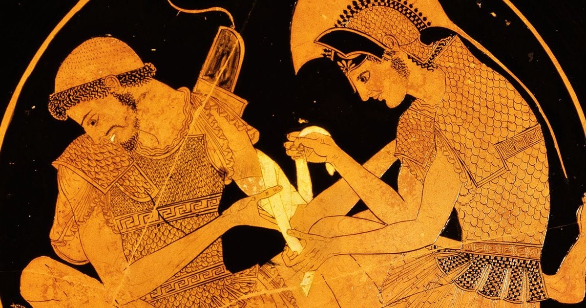
Ancient origins: early knowledge between religion and philosophy (3000 BCE – 200 CE)
The earliest traces of anatomical knowledge come from ancient civilizations, where the body is still a territory suspended between observation and symbol. In Egypt, certain practices (including those tied to ritual contexts) reveal basic notions about internal organs, yet representation remains largely symbolic more evocative than precise.
In Greece, medicine begins to take shape as a discipline: figures such as Hippocrates contribute to early medical writings and shift the focus toward rational explanation. Still, detailed anatomical imagery is rare. Tools are limited, shared methods are not yet established, and most importantly the cultural conditions for systematic observation are still fragile. Religious and social restrictions on dissection in multiple contexts significantly curb accuracy.
A first key point emerges here: medical-scientific communication grows not only through discoveries, but through the ability to see, verify, and compare.
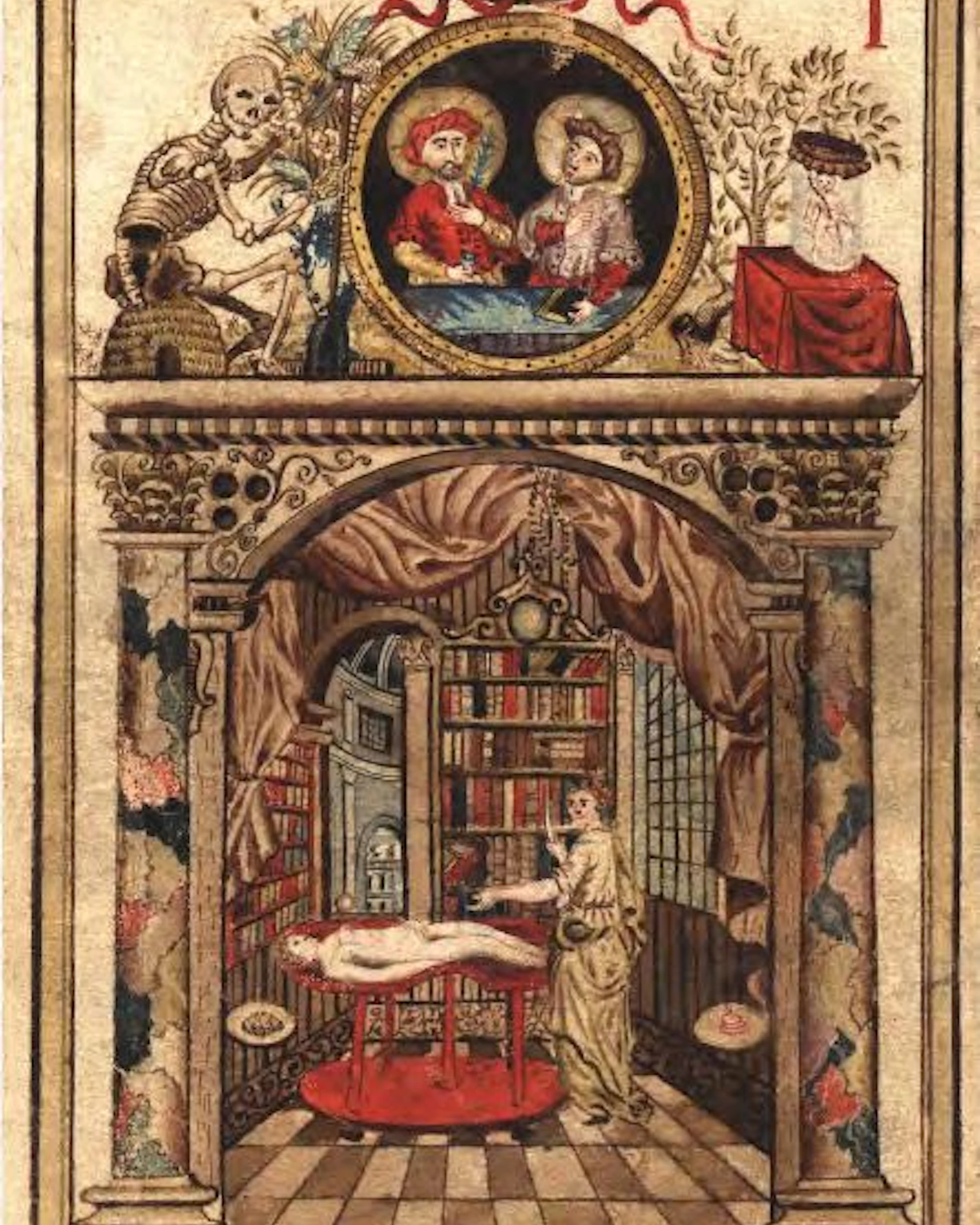
The Middle Ages: symbolism outweighs science (500 – 1400 CE)
In medieval Europe, anatomical progress slows. Medical knowledge relies heavily on the authority of Galen, preserved and expanded through Arabic scholarship, while anatomical illustration often remains inexact and shaped by symbolic visual language.
The body is represented within a framework that is more mystical than experimental: the human figure is “told” rather than observed. And what’s interesting is that, in this phase, images do not yet function as proof they are cultural translations of what is believed to be true.
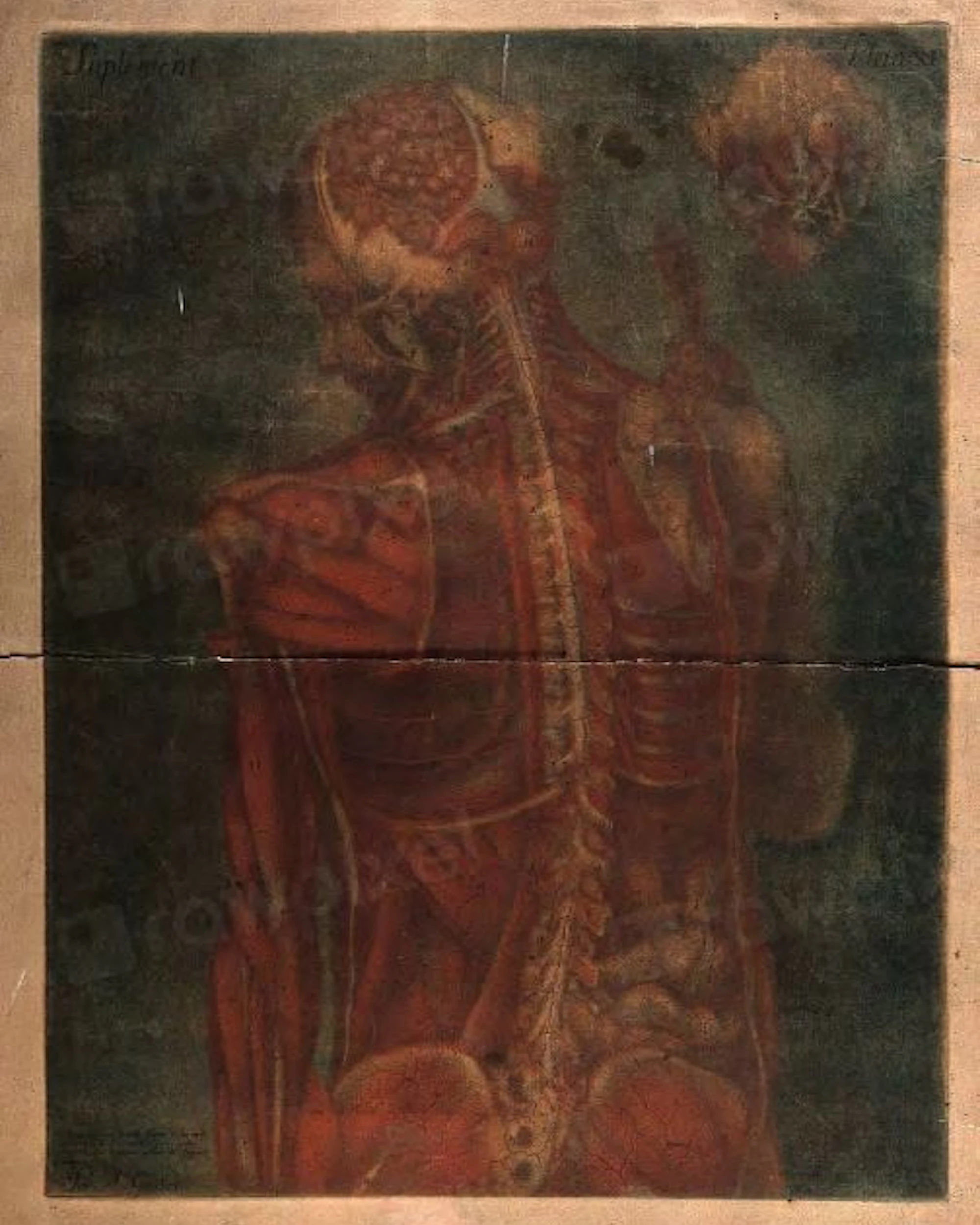
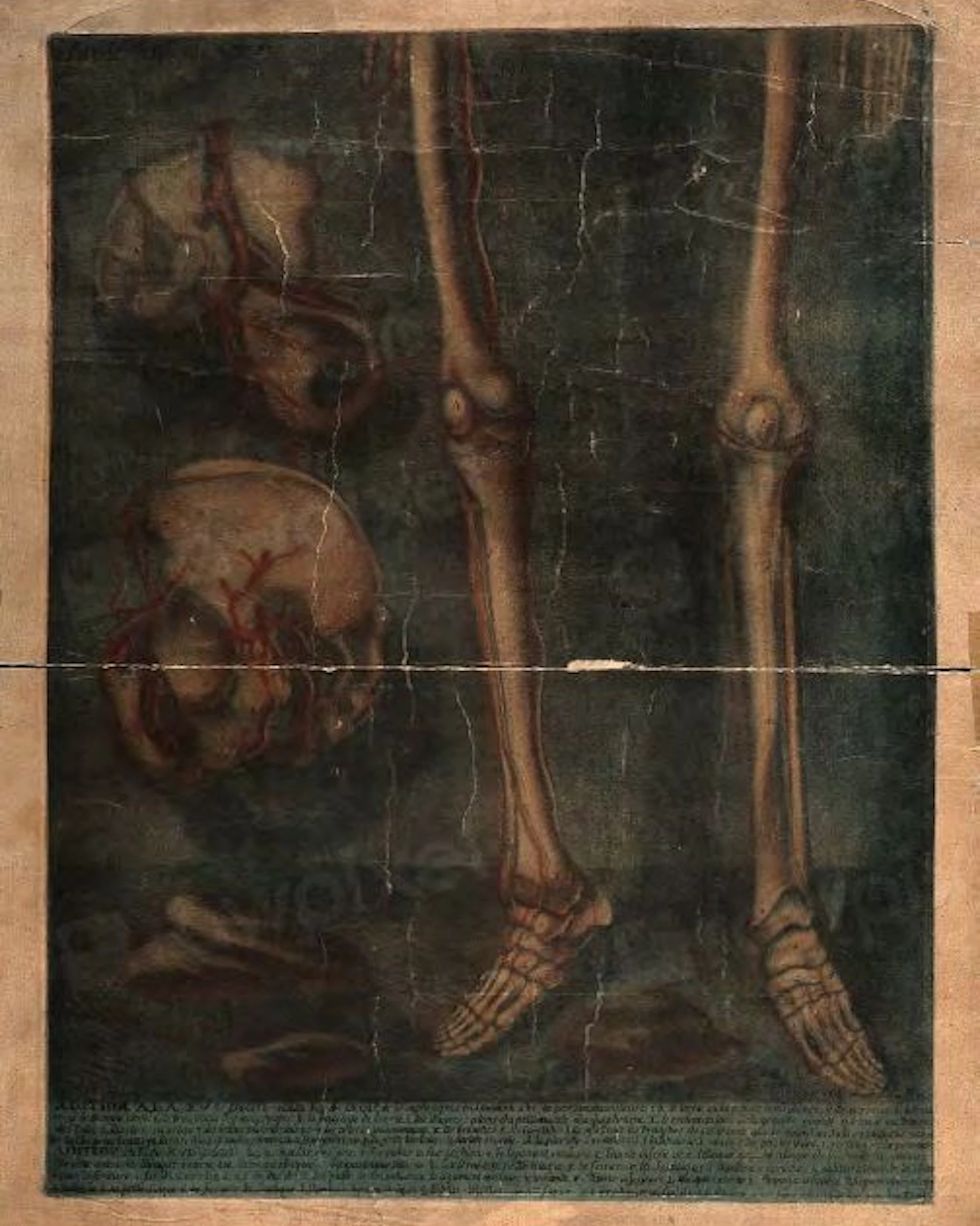
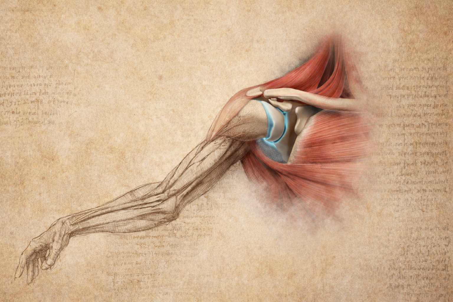
The Renaissance: a revolution in anatomical art (15th – 16th century)
The Renaissance changes the rules. Interest in science resurges, human dissection becomes possible again, and images finally become an investigative tool.
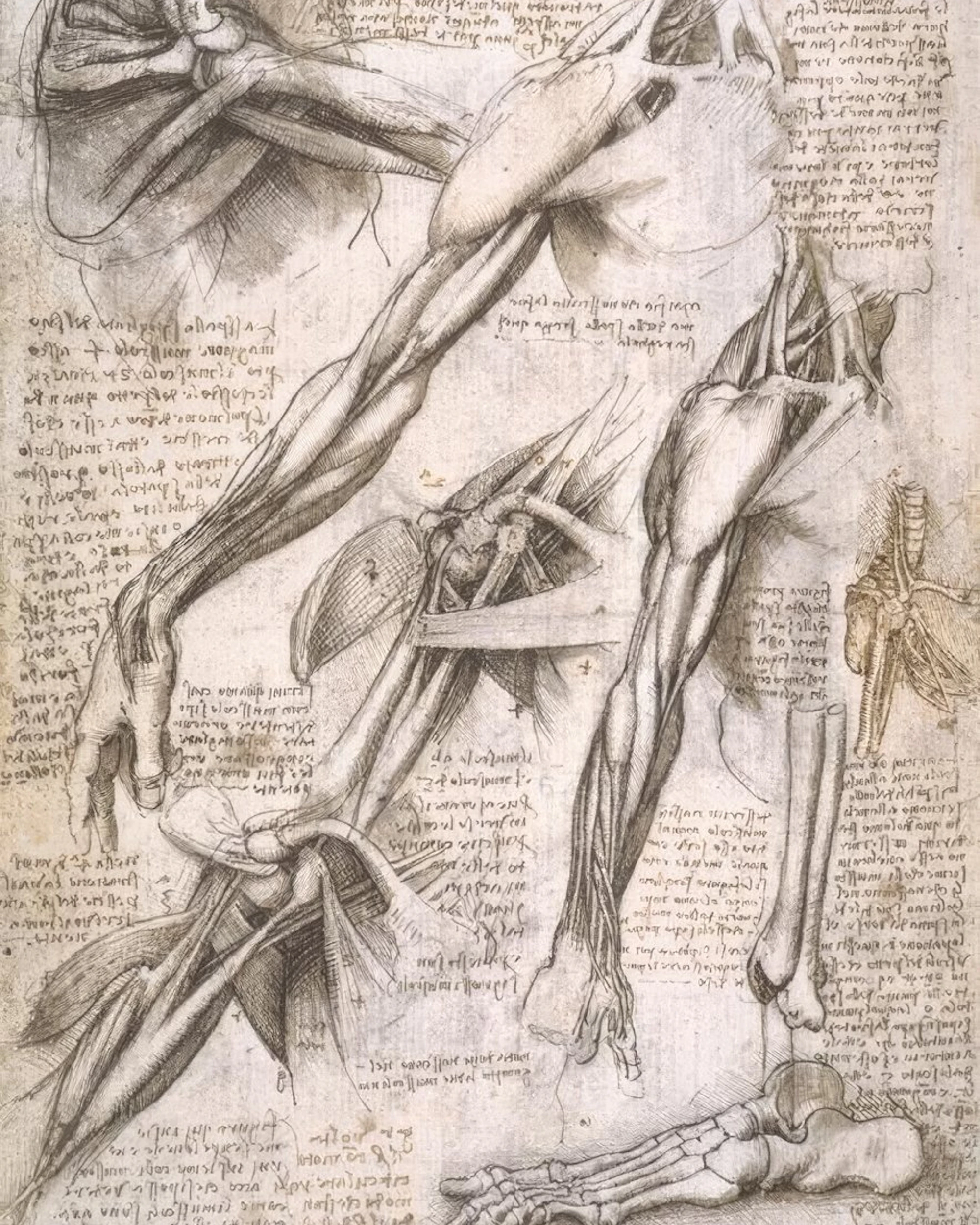
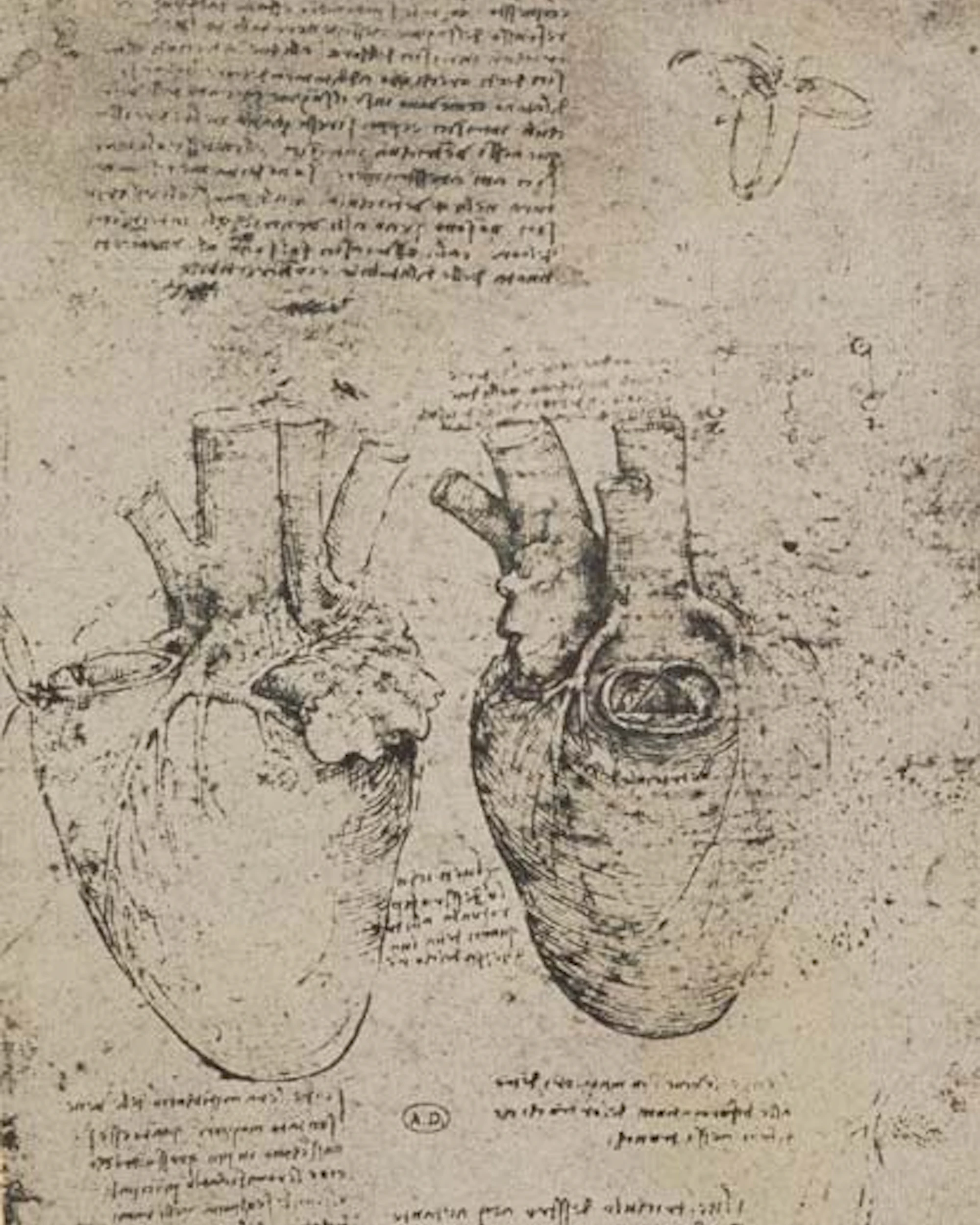
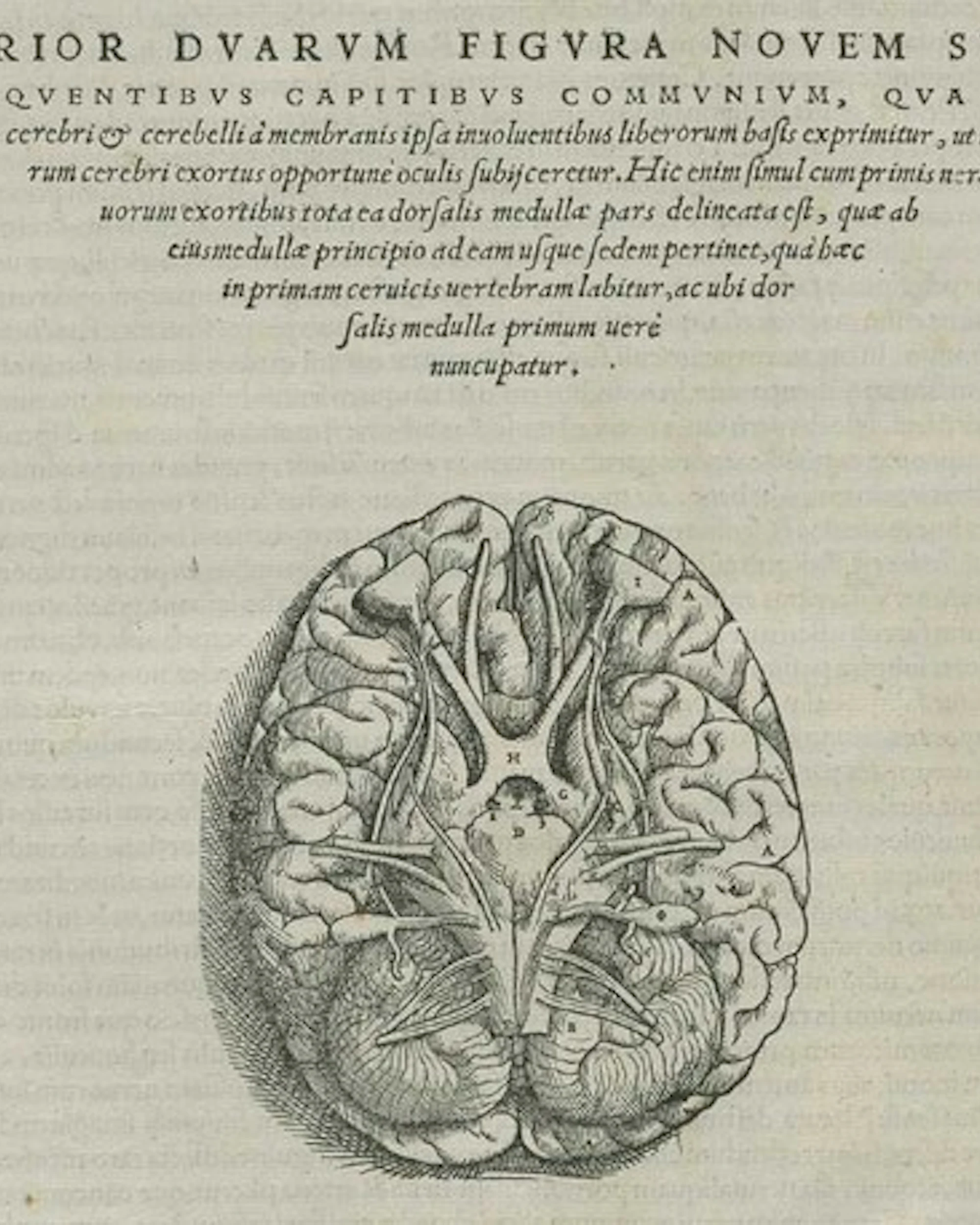
Figures like Leonardo da Vinci produce anatomically groundbreaking drawings remarkable for their detail and precision capable of capturing the complexity of muscles and organs. But the decisive turning point arrives with Andreas Vesalius: his De Humani Corporis Fabrica (1543) is considered a milestone because it is grounded in direct dissection, rather than relying solely on ancient texts.
Here, a fundamental shift takes place for medical-scientific communication: illustration stops being decoration and becomes visual rigor. Art and science begin working together toward a shared goal—making the body understandable through verifiable representation.
The Enlightenment and the rise of anatomical atlases (17th – 19th century)
After the Renaissance, scientific and technological progress accelerates and so does the way knowledge is shared. The spread of printing and the circulation of anatomical atlases support standardized education: knowledge no longer stays confined; it becomes more accessible and reproducible.
An iconic reference of this era is Gray’s Anatomy by Henry Gray (1858), still cited and used today, an work that makes anatomy more “navigable” through precise descriptions and detailed illustrations. In the 19th century, specialization increases (with examples such as William Hunter) and practical accuracy becomes a stronger priority.
Yet a crucial detail remains: the full correspondence with real anatomy the level of precision we now take for granted will truly mature only across the 20th and 21st centuries.
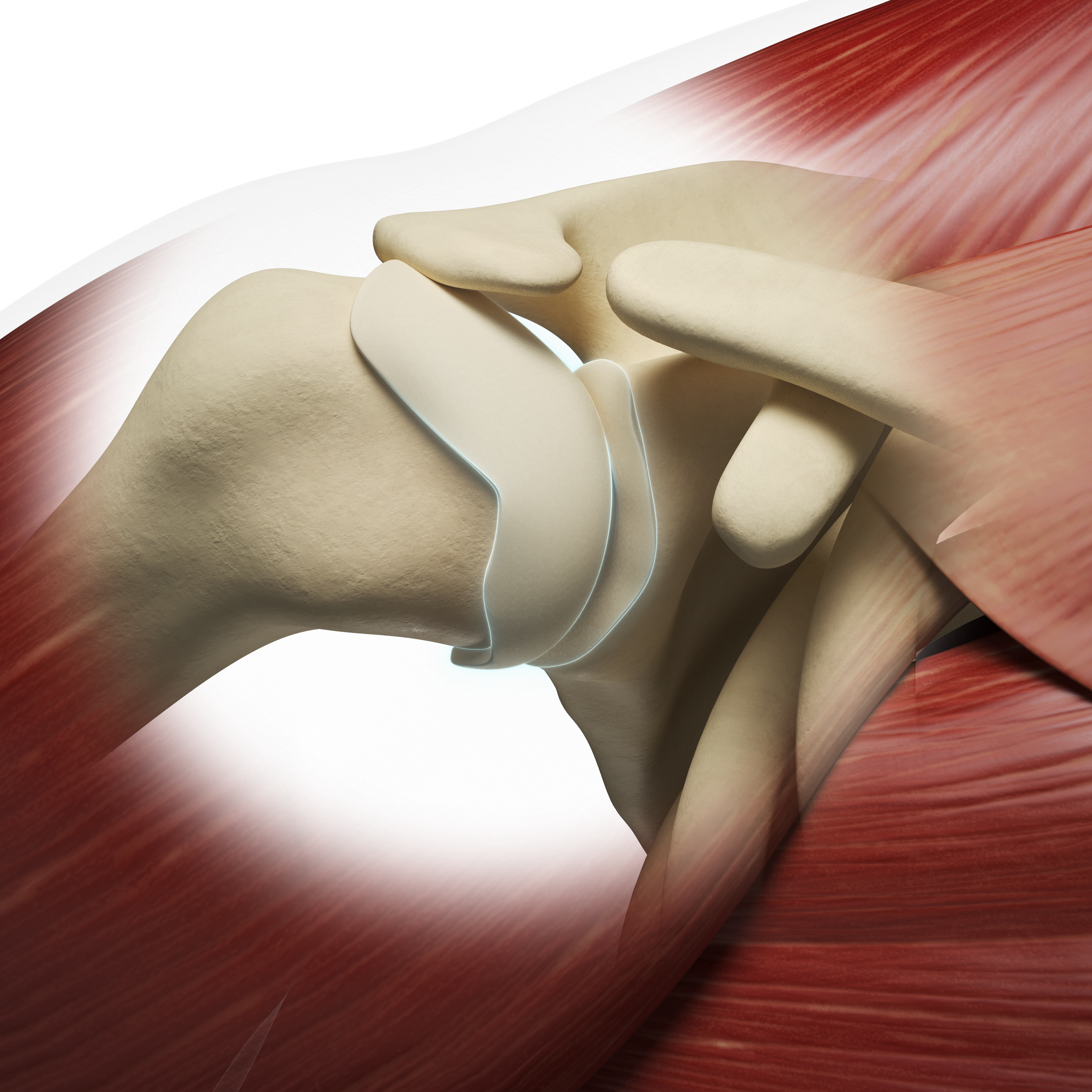
Modern times: photography, digital tools, and 3D modeling (20th century)
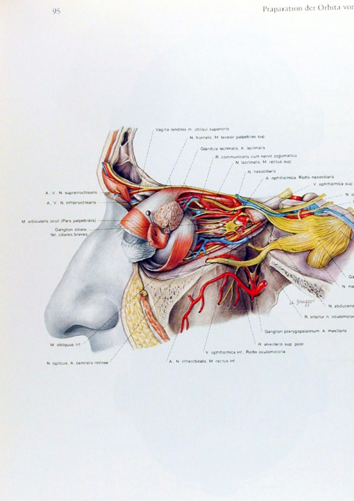
The 20th century introduces a radical transformation: photography and digital imaging deliver a level of precision and realism that traditional drawing could not reach. This is the moment when “seeing” becomes measurable and repeatable: no longer representation, but evidence.
Today, 3D modeling and advanced software translate anatomical knowledge into interactive, explorable models supported by medical imaging technologies for increasingly accurate visualization. These tools are not only educational: they enter surgical planning, training, and scientific communication both internally and externally.
And within this evolution, Artificial Intelligence is becoming part of the landscape as well enhancing the analysis of medical images and supporting faster, more scalable 3D model creation.